


- Gender
- Female
- Age
- 60
Description
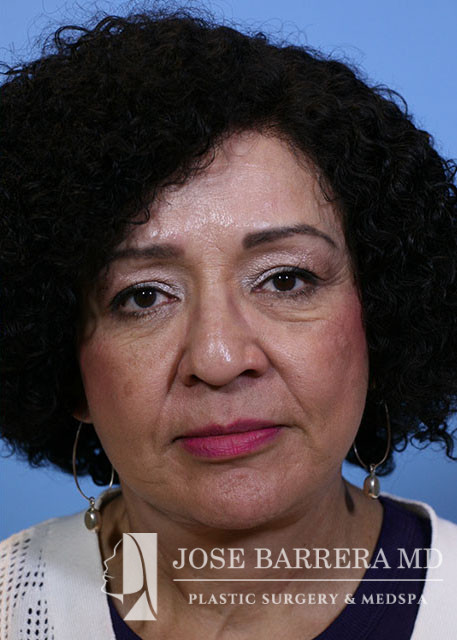
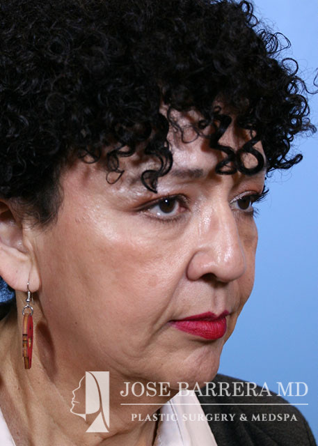
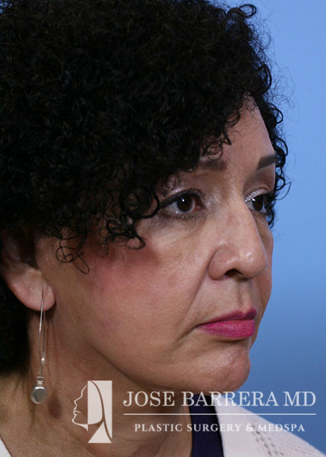
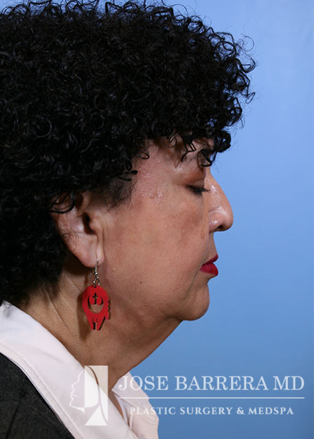
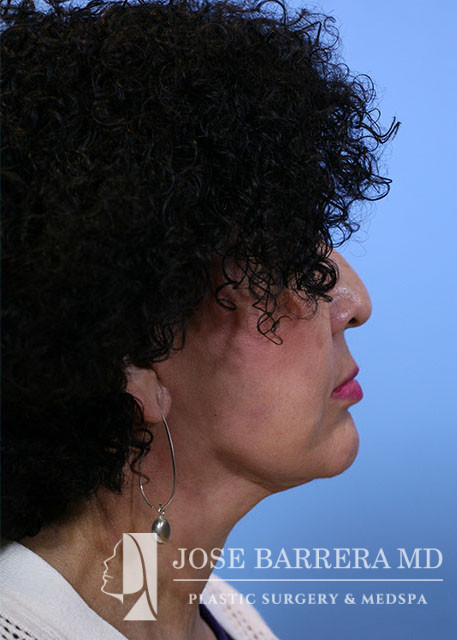
Operation: Deep Plane Facelift and Deep Necklift with Partial Digastric Muscle Resection Bilaterally
Indications: 59 year old female comes in for facial rhytidosis and neck banding.
Description of procedure:
Incisions for the facelift and necklift were previously marked out in the preoperative area. The preauricular portion of the incision was carried posterior to the tragus and the postauricular portion of the incision was carried on to the posterior auricle and into the hairline. These areas were locally anesthetized with tumescent mixture of 1% lidocaine, bupivacaine, epinephrine, normal saline, and tranexemic acid.
We then proceeded with the facelift and necklift. A submental incision made and a subcutaneous flap was developed extending from the inferior border of the mandible down to the thyroid cartilage and laterally to the angle of the mandible. Excess subcutaneous fat was sharply excised and the platysma was identified. We resected part of the anterior belly of the digastric muscle bilateral. Opposing platysma flaps were identified and dissected medially. A horizontal myotomy was made at the level of the hyoid. The platysma was then reapproximated using 4-0 PDS suture the in infra hyoid region. The suprahyoid platysma was the imbricated in the submental region with a running 4-0 PDS.
Next the right facelift incision was made. A subcutaneous flap was elevated, superficial to the great auricular nerve which was identified and preserved. The flap was connected to the plane developed in the neck. The subcutaneous flap was then elevated medially up to a line drawn from the lateral canthus to the angle of the mandible. At this point, the deep plane was entered. The SMAS was sharply incised and a subSMAS plane was developed. The zygomaticus major muscle was identified and care was taken to only dissect superficial to the muscle to protect branches of the facial nerve. The SMAS flap was further elevated medially and inferiorly to include the platysma. The flap was incised horizontally, parallel to the body of the mandible to allow for more mobility. Fat over the sternocleidomastoid was then excised to reduce bulk. The inferior portion of the SMAS flap was advanced posteriorly and secured to the mastoid periosteum with 3-0 PDS. Cervicocutaneous retaining ligament was also released and then secured posteriorly with 3-0 PDS. This created a well defined jawline. Fat was advanced along with this flap to allow for gonion augmentation. The superior portion of the flap was advanced superiorly and secured to the pre-auricular SMAS and fascia with 3-0 PDS. The area was inspected for any bleeding and careful hemostasis obtained. Excess skin was then carefully trimmed to ensure there was no tension on the final closure. The tragal skin was thinned. The same procedure was performed on the left face.