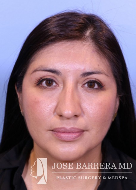
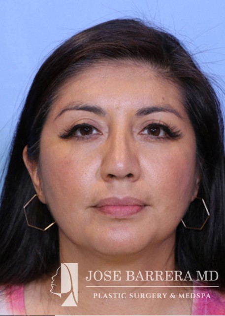
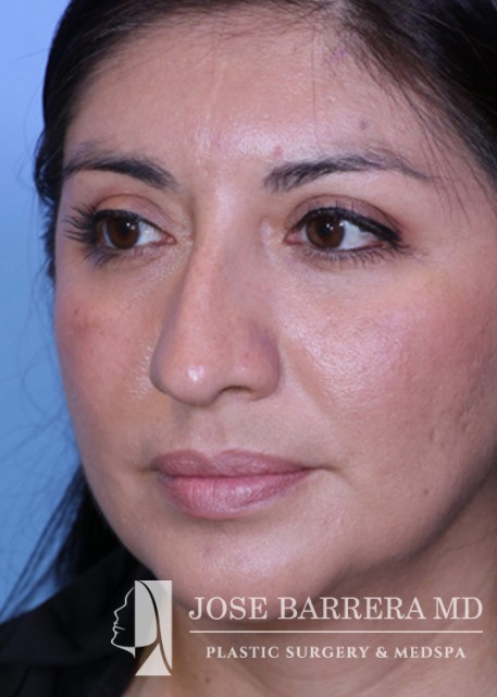

- Gender
- Female
Description
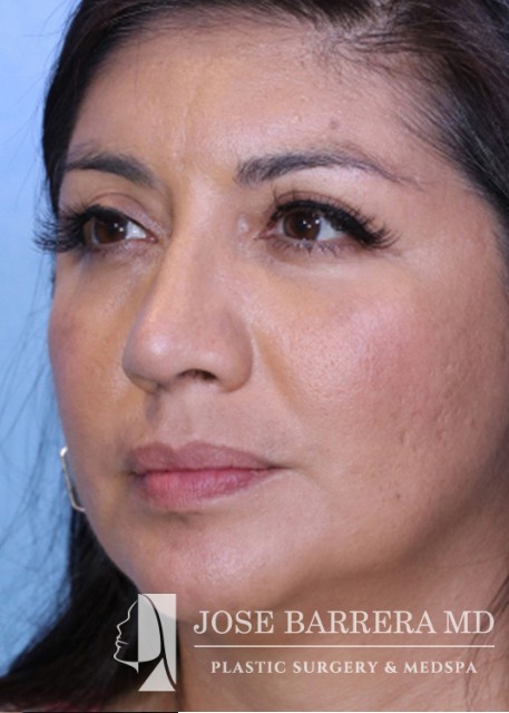
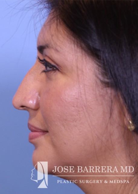
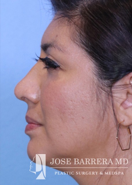
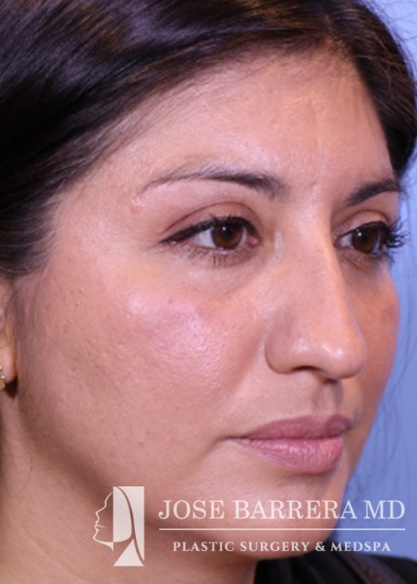
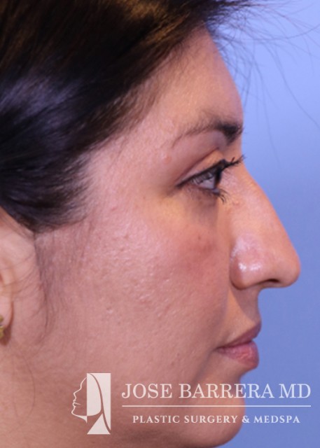
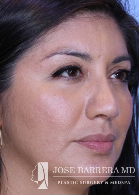
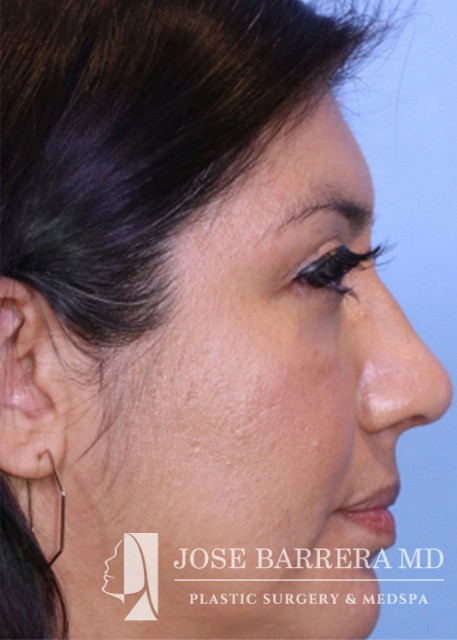
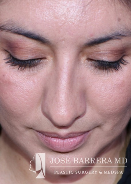
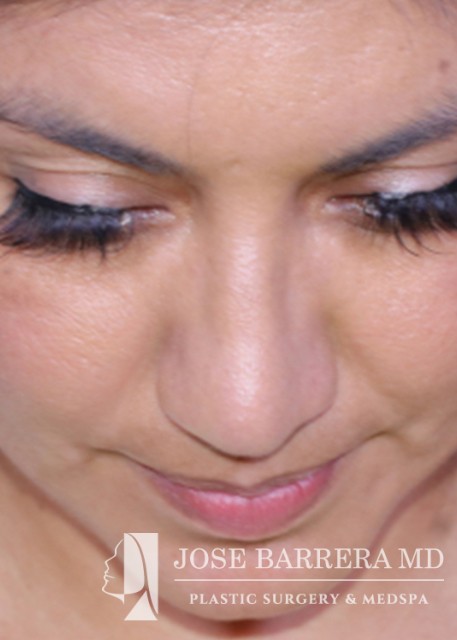
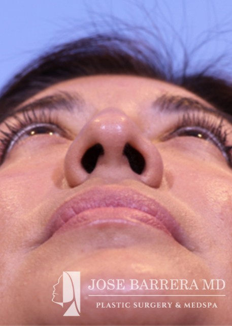
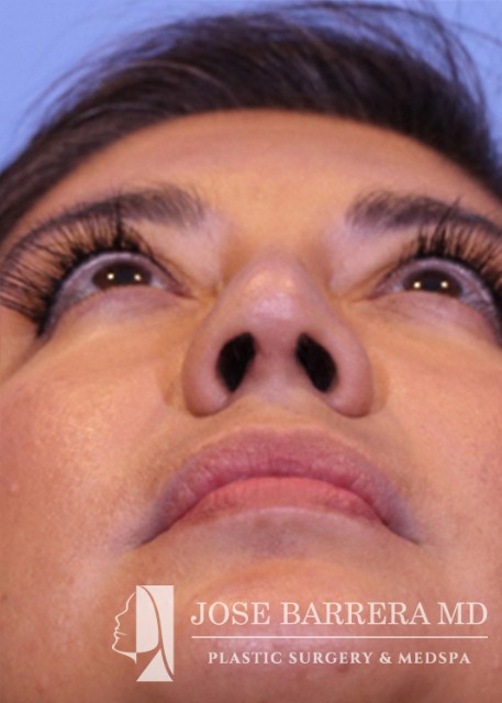
40-year-old female presents for initial consultation for rhinoplasty. She doesn’t like her dorsal hump or how her nose points down. An incision was then made along the pyriform aperture and the skin and soft tissue were elevated off the maxilla and nasal bones to prepare for osteotomies. The piezo saw was then used to make low-low-high osteotomies as well as a transverse osteotomy at the nasion. The dorsal roof remained intact. Small segments of nasal bone were removed at the pyriform aperture to allow for let down of the dorsal hump.The medial crura were separated sharply. The anterior septal angle was identified. A left mucoperichondrial flap was elevated. Dissection proceeded anteriorly around the caudal end of the septum allowing elevation of a mucoperichondrial flap on the right. Anterior upper lateral cartilages were released from the septum bilaterally.Next, the point of maximal dorsal convexity was identified and marked. A releasing vertical septal incision was made inferior to the point of maximal dorsal convexity. A modified Z plasty dorsal septum cut was made that connected to the vertical releasing incision. An osteotomy was also made through the perpendicular plate of the ethmoid bone that connected to the transverse osteotomy. At this point, the nasal dorsum was fully let down. The anterior wedge of cartilage was overlapped on the left side of the native septum. Multiple 5-0 prolenes were placed around the mobilized dorsal septum to hold the let down dorsal hump in position.Inferior septal cartilage was harvested in one piece with care taken to maintain greater than 1 cm T strut. The anterior upper lateral cartilages were further released from the septum. A septal extension graft was fashioned from the harvested septal cartilage and secured to the left side of the native septum with 5-0 prolene. A short spreader graft was fashioned and placed on the left for further support. 5-0 prolene dome spanning and lateral crural tensioning sutures were placed to create new domes and increase rotation, securing the lower lateral cartilages to the septal extension graft. The skin was elevated off the medial crura. A tongue-and-groove procedure was performed and the medial crura were sutured to the caudal septum with 5-0 prolene. Medial crural fixation sutures were placed to secure the medial crura together and narrow the columella.