


- Gender
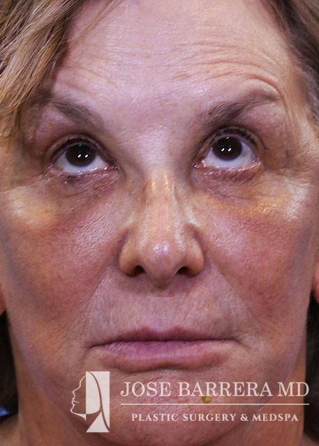
- Female
- Age
- 69
Description
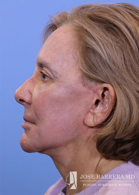
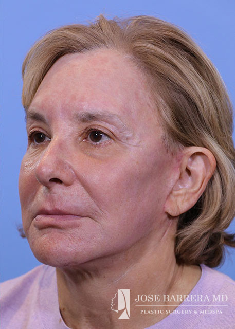
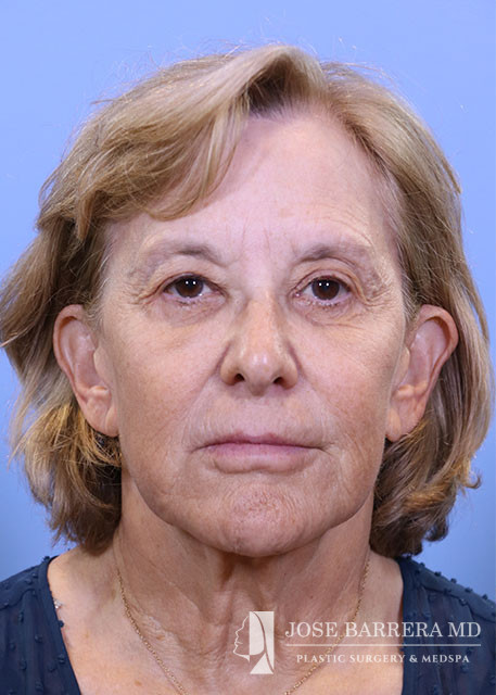
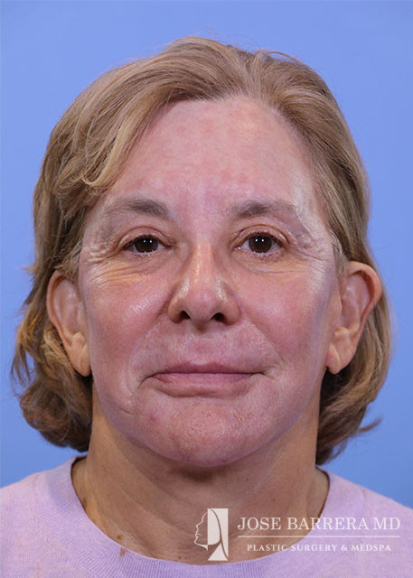
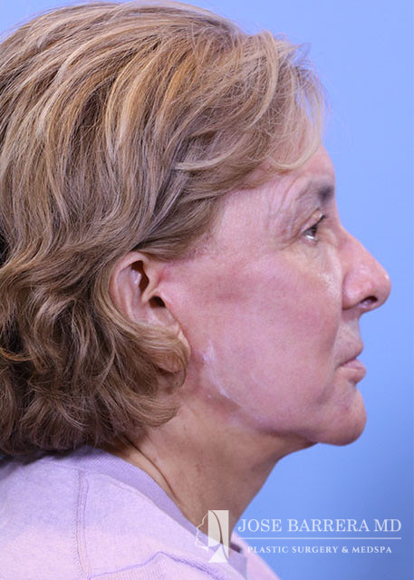
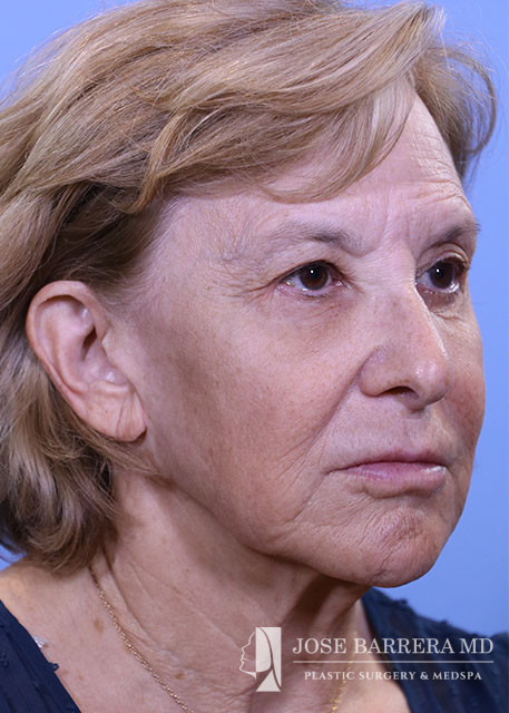
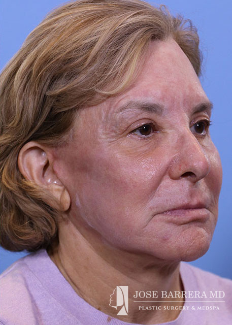
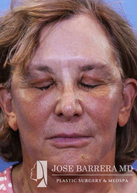
FINDINGS: Deep plane facelift/neck lift, endoscopic brow lift with endotines, upper blepharoplasty, lower blepharoplasty with resection of lateral fat pads.
INDICATIONS: Brow ptosis, dermatochalasis, unacceptable cosmetic appearance
DESCRIPTION OF PROCEDURE: The patient was brought to the OR and identified by name and date of birth. IV anesthesia was given and oxygen administered via LMA.
We then proceeded with upper blepharoplasty. 8 mm of upper eyelid skin on each side was excised. Hemostasis obtained. The incision was closed with a subcuticular 6-0 prolene. The ends of the prolene were secured in place with steri strips.
We then proceeded with lower blepharoplasty via transconjunctival approach. The area was anesthetized with 1% lidocaine with 1:100,000 epinephrine. The right side was addressed first. Incision made through conjunctiva and dissection proceeded in a preseptal plane. The periosteum overlying the orbital rim was incised. Periosteum elevated off the orbital rim. Hemostasis obtained. The orbital septum was incised and herniating fat from the lateral fat pad was excised with bipolar cautery. The same complete procedure was then performed on the the left lower eyelid.
The forehead was then addressed. An incision made down through periosteum to the bone and planned skin was excised. The flap was elevated off the bone down to the superior orbital rim. The supraorbital and supratrochlear nerves were identified and preserved. Galea in the area around the nerves was released using an elevator and the retro-orbicularis oculi fat was identified. Through the lateral most incisions, the temporalis muscle was identified. Dissection carried out in a plane superficial to the true temporal fascia. The conjoined tendon and the arcus marginalis were released bilaterally.
Through each paramedian incision, a hole was drilled with a hand drill. Endotine implants were then placed. The forehead flap was advanced superiorly and secured on the endotines. This allowed for appropriate elevation of the brow. All five scalp incisions were all closed with 4-0 PDS deep and staples on skin.
We then proceeded with the face and necklift. Incisions for the facelift and necklift were previously marked out in the preoperative area. The preauricular portion of the incision was carried posterior to the tragus and the postauricular portion of the incision was carried on to the posterior auricle and into the hairline. These areas were locally anesthetized with tumescent mixture of 1% lidocaine, bupivacaine, epinephrine, and normal saline.
We then proceeded with the facelift and necklift. A submental incision made and a subcutaneous flap was developed extending from the inferior border of the mandible down to the thyroid cartilage and laterally to the angle of the mandible. Excess subcutaneous fat was sharply excised and the platysma was identified. Opposing platysmal flaps were identified and dissected medially. A right tension band was incised and released. Separately there was prolapse of the left submandibular gland that was removed with bipolar cautery. The platysma was then reapproximated using 4-0 PDS suture the in infra hyoid region. The suprahyoid platysma was the imbricated in the submental region with a interrupted 4-0 PDS.
Next the right facelift incision was made. A subcutaneous flap was elevated, superficial to the great auricular nerve which was identified and preserved. The flap was connected to the plane developed in the neck. The subcutaneous flap was then elevated medially up to a line drawn from the lateral canthus to the angle of the mandible. At this point, the deep plane was entered. The SMAS was sharply incised and a subSMAS plane was developed. The zygomaticus major muscle was identified and care was taken to only dissect superficial to the muscle to protect branches of the facial nerve. The SMAS flap was further elevated medially and inferiorly to include the platysma. The flap was incised horizontally, parallel to the body of the mandible to allow for more mobility. Fat over the sternocleidomastoid was then excised to reduce bulk. The inferior portion of the SMAS flap was advanced posteriorly and secured to the mastoid periosteum with 3-0 PDS. Cervicocutaneous retaining ligament was also released and then secured posteriorly with 3-0 PDS. This created a well defined jawline. Fat was advanced along with this flap to allow for gonion augmentation. The superior portion of the flap was advanced superiorly and secured to the pre-auricular SMAS and fascia with 3-0 PDS. The area was inspected for any bleeding and careful hemostasis obtained. Excess skin was then carefully trimmed to ensure there was no tension on the final closure. The tragal skin was thinned. The same procedure was performed on the left face.